Always an AMDG Dentist near you

Radiological examination
These are made with the help of a film or a digital sensor and are divided into 2 groups:
- Intra oral: the film is placed inside the mouth
- Extra oral: the film is placed outside the mouth.
Intraoral radiographs are the most common:

Bitewings
the film or sensor is placed just behind the crowns of the molars and premolars. This x-ray allows the detection of interdental cavities (caries) and allows an evaluation of the bone structure close to the crowns.
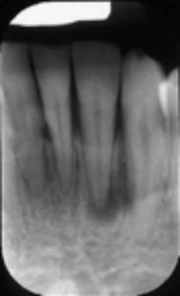
Apical x-rays
the film or sensor is placed along the axis of the teeth next to the tongue or palate. This x-ray allows the detection of bone lesions (cysts or apical lesions).
Extra oral radiographs:
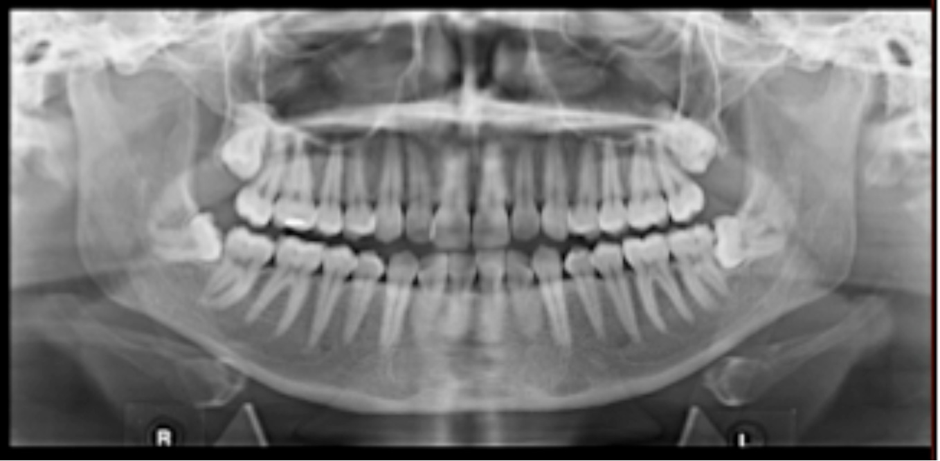
Panoramique (OPT)
gives an overall view of the teeth and supporting bone. It is less precise than the intraoral radiographs, particularly for detecting caries.

Lateral head-film
this x-ray is an essential part of an orthodontic documentation; its analysis determines the horizontal and vertical position of the maxillae, and the axis of the incisors. It also allows a follow-up of the patients growth pattern and coarse of treatment.

Cone beam CT
this is the most precise but also the exam that emits the most radiation, although less than a medical scanner. It is performed if a three-dimensional vision is necessary, usually in surgery or implantology.
A specialist in dentofacial imaging makes the realization and interpretation of this image in a radiological institution.